Food Science and Preservation
The Korean Society of Food Preservation
Research Article

TNF-α/IFN-γ로 자극된 HaCaT 및 IgE로 감작된 RBL-2H3 세포에서 닥나무 가지 추출물의 알러지 염증반응 억제 효과

배원빈1,https://orcid.org/0009-0009-5733-5972, 김은혜2https://orcid.org/0009-0005-0793-1259, 김민주1https://orcid.org/0009-0005-7918-9428, 양선아1,,*;https://orcid.org/0000-0002-6692-1844
Won-Bin Bae1,https://orcid.org/0009-0009-5733-5972, Eun-Hye Kim2https://orcid.org/0009-0005-0793-1259, Min-Ju Kim1https://orcid.org/0009-0005-7918-9428, Seun-Ah Yang1,,*;https://orcid.org/0000-0002-6692-1844
1계명대학교 식품가공학과
2금오공과대학교 응용화학과
1Department of Food Science and Technology, Keimyung University, Daegu 42601, Korea
2Department of Applied Chemistry, Kumoh National Institute of Technology, Gumi 39177, Korea
*Corresponding author Seun-Ah Yang, Tel: +82-53-580-5117, E-mail: seunahy@kmu.ac.kr

These authors contributed equally to this study.

Citation: Bae WB, Kim EH, Kim MJ, Yang SA. Inhibitory effects of Broussonetia kazinoki twig extract on allergic inflammatory reactions in TNF-α/IFN-γ-stimulated HaCaT and IgE-sensitized RBL-2H3 cells Food Sci. Preserv., 31(2), 307-314 (2024)

Copyright © The Korean Society of Food Preservation. This is an Open Access article distributed under the terms of the Creative Commons Attribution Non-Commercial License (http://creativecommons.org/licenses/by-nc/4.0) which permits unrestricted non-commercial use, distribution, and reproduction in any medium, provided the original work is properly cited.

Received: Jan 16, 2024; Revised: Feb 16, 2024; Accepted: Feb 19, 2024

Published Online: Apr 30, 2024

Abstract

Broussonetia kazinoki twig extract (BKT) is recognized for its antioxidant and anti-cancer effects and natural whitening properties. So, it is used as a raw material for cosmetics. B. kazinoki twig is also an edible raw material. B. kazinoki has been used in Asia for paper production and oriental medicine, has anti-diabetic effects, and contains various flavonoids and alkaloids. In this study, to evaluate the efficacy of BKT on allergic skin inflammatory responses, we investigated its effects on factors related to skin inflammation in HaCaT keratinocytes and allergic responses in RBL-2H3 cells. There was no cytotoxicity of the 70% ethanol extract against HaCaT and RBL-2H3 cells. In HaCaT cells, stimulation with tumor necrosis factor-alpha (TNF-α) and interferon-gamma (IFN-γ) increased the production of several chemokines, including thymus and activation-regulated chemokine (TARC), macrophage-derived chemokine (MDC), and regulated on activation, normal T cell expressed and secreted (RANTES). However, it was observed that this elevation was notably mitigated in a concentration-dependent manner upon treatment with BKT. Furthermore, BKT treatment demonstrated a significant reduction of β-hexosaminidase and inflammatory cytokines TNF-α and IL-4 in IgE-sensitized RBL-2H3 cells. Thus, it is expected that BKT can be used as a natural cosmetic and food ingredient that effectively suppresses allergic inflammatory reactions.

Keywords: Broussonetia kazinoki; anti-inflammation; anti-allergy; HaCaT keratinocytes; RBL-2H3 cells

1. 서론

현대 사회에서 피부염증과 알러지 반응은 인간 건강과 삶의 질에 영향을 미치는 심각한 문제로 간주되며, 이러한 문제는 음식, 환경 오염, 알레르겐 및 박테리아 노출, 유전적 요인 등 다양한 원인으로 인해 증가하고 있는 실정이다(Lee 등, 2014). 이러한 피부염증과 알러지 반응은 피부각질세포 및 면역세포의 활성화와 상호작용에 의해 유발되며, 관련된 세포 및 분자의 활동 조절은 염증과 알러지의 진행을 조절하는 데 중요한 역할을 한다(Chung 등, 2011; Kim 등, 2023). 피부 내 각질형성세포인 keratinocyte는 표피층에 존재하는 주요 구성 세포로, 세포 분화를 통해 피부 장벽을 형성하는 역할을 한다(Oh와 Jang, 2015). 이러한 피부각질세포가 수많은 항원과 접촉하고 스트레스 환경에 노출되면 다양한 케모카인(chemokine)과 사이토카인(cytokine) 등 염증 매개 인자를 생성한다(Jo, 2022). 장기적인 피부 손상으로 케모카인과 사이토카인이 지속적으로 생성되면 피부염증 및 건조를 유발하여 피부염, 건선, 아토피 피부염(atopic dermatis, AD) 등의 피부질환이 유도된다(Hudson, 2006). 알러지에 의해 발생하는 피부염증 질환 중 하나인 급성 AD는 T-helper 2-type 세포(Th2)의 피부 병변으로의 침윤 및 Th2 관련 사이토카인인 인터루킨(Interleukin, IL)-4, IL-5 및 IL-13의 분비 증가로 정의된다(Lee 등, 2010). 대부분의 AD 환자는 Th1 세포 수가 감소하고 Th2 반응에 대응할 수 있는 인터페론(IFN-)과 같은 Th1 사이토카인의 농도가 낮으며(Lee 등, 2010), 초기 면역글로불린 E (immunoglobulin E, IgE) 생산 및 이후 알레르겐/IgE 반응과 관련이 있을 가능성이 높은 것으로 알려져 있다(Bergmann 등, 1998). Th2 세포는 Thymus and activation-regulated chemokine (TARC/CCL17)과 macrophage-derived chemokine (MDC/CCL22)에 의해 유도되며, TARC 및 MDC는 AD와 같은 일부 피부 질환의 발병에 중요한 역할을 하는 것으로 사료된다. 다수의 연구에서, AD 환자의 혈청 중 TARC와 MDC 케모카인 수준이 현저하게 상승하며, 이러한 케모카인 수준의 상승과 환자의 질병 심각도 간에 상관관계가 있는 것으로 확인되었다(Furukawa 등, 2004; Hashimoto 등, 2006). 더불어, AD와 관련이 있는 또 다른 케모카인인 Regulated on activation, normal T cell expressed and secreted (RANTES/CCL5)도 AD와 관련이 있는 것으로 알려져 있다(Homey와 Zlotnik, 1999). RANTES/CCL5는 AD 환자의 피부에서 높은 농도로 검출되었으며, 이 케모카인의 주요 공급원은 진피 섬유아세포로 확인되었다(Schroder 등, 1996).

알러지 반응은 항원의 결합에 의해 유발되며, 비만세포(mast cell)와 호염기구 표면의 고친화성 immunoglobulin E (IgE) 수용체인 Fc∊RI에 결합한다. 이 반응은 알러지 관련 사이토카인 합성 및 분비, 세포 내 반응성 산소종(ROS) 생성 및 항원 자극 탈과립화로 이어진다(Itoh 등, 2011). 또한, histamine과 β-hexosaminidase 등이 방출되고, 염증성 사이토카인[rat tumor necrosis factor (TNF-α), interleukin-4 (IL-4) 등]과 염증 매개체를 방출하여 알러지 질환 발병에 중요한 역할을 한다(Chung 등, 2013; Sicherer과 Sampson, 2018).

닥나무(Broussonetia kazinoki)는 아시아 국가에서 오랫동안 종이 제조 및 한의학적 용도로 사용되어 왔다. 닥나무 추출물은 한의학에서 화상, 여드름 등의 피부질환에도 사용되며 항당뇨, 항고혈당 효능이 있고(Cha 등, 2008), 티로시나제 및 멜라닌 합성(Hwang과 Lee, 2007)과 산화질소 과잉생산을 억제(Ryu 등, 2003)하는 효능이 보고되었다. 알러지 반응에서 닥나무 잎과 가지에는 broussonol A, B, C, D, 그리고 E (Zhang 등, 2001) 와 같은 플라보노이드와 broussonetines A, B, W, X, 그리고 J1 (Tsukamoto 등, 2001)을 포함한 알칼로이드를 함유하는 것으로 알려져 있고, 닥나무 줄기 및 심재의 항염증, 항알러지 효능이 보고되었으나(Lee 등, 2008; Lee, 등, 2010), 현재까지 닥나무 가지 추출물의 항피부염증, 항알러지 효능은 보고되지 않았다. 따라서 본 연구에서는 HaCaT (human keratinocyte cell type) 세포에서 TARC, MDC, RANTES 생성량 변화를 통한 닥나무 가지 추출물의 항염증 효능과, RBL-2H3 (rat basophilic leukemia cell line) 세포에서 β-hexosaminidase 방출량, TNF-α, IL-4 생성량 변화를 통한 항알러지 효능을 평가하고자 하였다.

2. 재료 및 방법

2.1. 닥나무 가지 추출물 제조

건조된 닥나무 가지는 청명약초(Chungju-si, Korea)에서 구입하여 사용하였다. 닥나무 가지는 5-10 mm의 크기로 분쇄하였다. 추출물 제조를 위하여 분쇄된 닥나무 가지 1 kg에 20 L의 70% 에탄올을 가하고, 80°C에서 6시간 동안 추출을 하였다. Filter paper (Advantec, Tokyo, Japan)를 이용하여 추출액을 여과한 후, 회전 감압농축기(Tokyo Rikakikai, Tokyo, Japan)로 농축하고 동결건조(FDU-2100, EYELA, Tokyo, Japan)하여 제조한 분말을 시료로 사용하였다.

2.2. HaCaT 피부각질세포 및 RBL-2H3 세포 배양

세포 배양은 Lee 등(2020)의 실험 방법을 이용하였다. 실험에 사용된 인간 유래 HaCaT 피부각질세포는 cell line service (CLS, Eppelheim, Germany), RBL-2H3 세포는 American Type Culture Collection (ATCC, Rockville, ND, USA)로부터 분양 받아 사용하였으며, HaCaT 피부각질세포는 10% fetal bovine serum, 1% penicillin-streptomycin을 첨가한 Dulbecco’s modified Eagle’s Medium (DMEM) 배지, RBL-2H3 세포는 10% fetal bovine serum, 1% penicillin-streptomycin을 첨가한 Minimum Essential Medium (MEM) 배지를 사용하여 5% CO2, 37°C 배양기에서 배양하여 실험에 사용하였다.

2.3. HaCaT 피부각질세포 및 RBL-2H3 세포 생존율 측정

닥나무 가지 추출물의 HaCaT 피부각질세포와 RBL-2H3 세포에 대한 세포 독성을 Ha 등(2020), Lee 등(2023)의 방법을 참고하였으며, 3-(4,5-dimethylthiazol-2-yl)-2,5-diphenyl-tetrazolium bromide (MTT) assay를 이용하여 평가하였다. HaCaT 피부각질세포는 2×10⁵ cells/well로 96 well plate에 분주하고 24시간 동안 배양 후, 10 ng/mL의 TNF-α와 interferon-γ (IFN-γ)가 포함된 DMEM 180 μL와 시료 20 μL를 첨가하고 24시간 재배양하였다. RBL-2H3 세포는 3×10⁵ cells/well의 비율로 24 well plate에 분주하고 24시간 동안 배양 및 anti-dinitrophenyl-immunoglobulin E (anti-DNP-IgE)를 함유한 MEM 배지를 사용하여 24 well plate에 세포를 분주하고 24시간 동안 감작하고 시료 처리 후 5 μg/mL MTT가 포함된 DMEM 배지로 교체하고 4시간 동안 배양하였다. 이후 상등액을 제거하고 DMSO를 넣어 형성된 formazan 결정을 용해하고 550 nm에서 흡광도를 측정하였다.

2.4. HaCaT 피부각질세포를 이용한 항염증 활성 측정

HaCaT 피부각질세포를 이용한 항염증 활성은 Lee 등(2014)의 방법을 이용하여 측정하였다. HaCaT 피부각질세포를 2×10⁵ cells/well로 96 well plate에 분주하고 24시간 동안 배양하였다. 이후 상등액을 모두 제거하고 10 ng/mL의 TNF-α와 interferon-γ (IFN-γ)가 포함된 DMEM 180 μL와 시료 또는 silymarin (positive control) 20 μL를 첨가하고 24시간 재배양하였다. TNF-α/interferon-γ (T+I) 10 ng/mL로 유도된 HaCaT 피부각질세포의 닥나무 가지 추출물에 의한 독성은 MTT assay를 이용하여 평가하였다. 또한, 배양 후 상등액에 존재하는 TARC, MDC, RANTES 함량은 R&D systems에서 구입한 Quantikine ELISA Kit를 이용하여 제조사 지침에 따라 측정하였다.

2.5. RBL-2H3 세포를 이용한 β-hexosaminidase 저해 활성 측정

닥나무 추출물의 항알러지 효능을 확인하기 위하여 β-hexosaminidase 방출량을 측정하였다. β-hexosaminidase 방출량 측정은 Lee 등(2020)의 실험 방법을 이용하였다. RBL-2H3 세포를 3×10⁵ cells/well의 비율로 24 well plate에 분주하고 24시간 동안 배양하였다. 이후 각 세포들을 1X PBS를 이용하여 세척하고 0.05 μg/mL의 anti-dinitrophenyl-immunoglobulin E (anti-DNP-IgE)를 함유한 MEM 배지를 사용하여 24 well plate에 세포를 분주하고 24시간 동안 감작하였다. 감작된 RBL-2H3 세포를 1% FBS를 포함하는 MEM 배지로 2회 세척한 뒤, 160 μL 배지로 교체하고 20분간 배양하였다. 닥나무 추출물을 농도별로 희석하여 세포에 처리하고 1시간 배양 후, 0.1 μg/mL DNP-HSA (Human serum albumin)를 처리하고 4시간 동안 배양 후 상등액을 원심분리(4°C, 13,000 rpm, 10분)하였다. 원심분리 후 남은 상층액 25 μL를 96 well plate에 옮기고, 0.1 M sodium citrate buffer (pH 4.5)에 10 mM로 희석되어 있는 4-p-nitrophenyl-N-acetyl-β-D-glucosaminide을 50 μL 혼합하여 1시간 동안 37°C에서 반응하였다. 0.1 M carbonate buffer (pH 10.0)를 첨가하여 반응을 종결시키고 405 nm에서의 흡광도를 microplate reader (Bio-Tek Instruments, Winooski, VT, USA)를 이용하여 측정하였다.

2.6. Cytokine 생성량 측정

세포배양액 내의 rat TNF-α (Abcam), rat IL-4 (Abcam, Cambridge, UK) 함량은 Chung 등(2020)의 방법을 참고하였으며, ELISA kit를 이용하여 제조사 지침에 따라 microplate reader로 측정하였다.

2.7. 통계처리

본 실험 결과들은 3회 반복 실험을 수행하고, 평균과 표준편차(mean±SD)로 나타내었다. 각 데이터는 statistical package for social sciences (SPSS 29.0, 2.0, IBM Corp., Armonk, NY, USA) software를 이용하여 one-way ANOVA를 실시한 후 유의성이 나타난 경우 집단 간 유의적인 차이를 Duncan의 사후검정(p<0.05)으로 집단 간의 차이를 검정하였다.

3. 결과 및 고찰

3.1. 닥나무 가지 추출물의 HaCaT cell에 대한 세포 독성

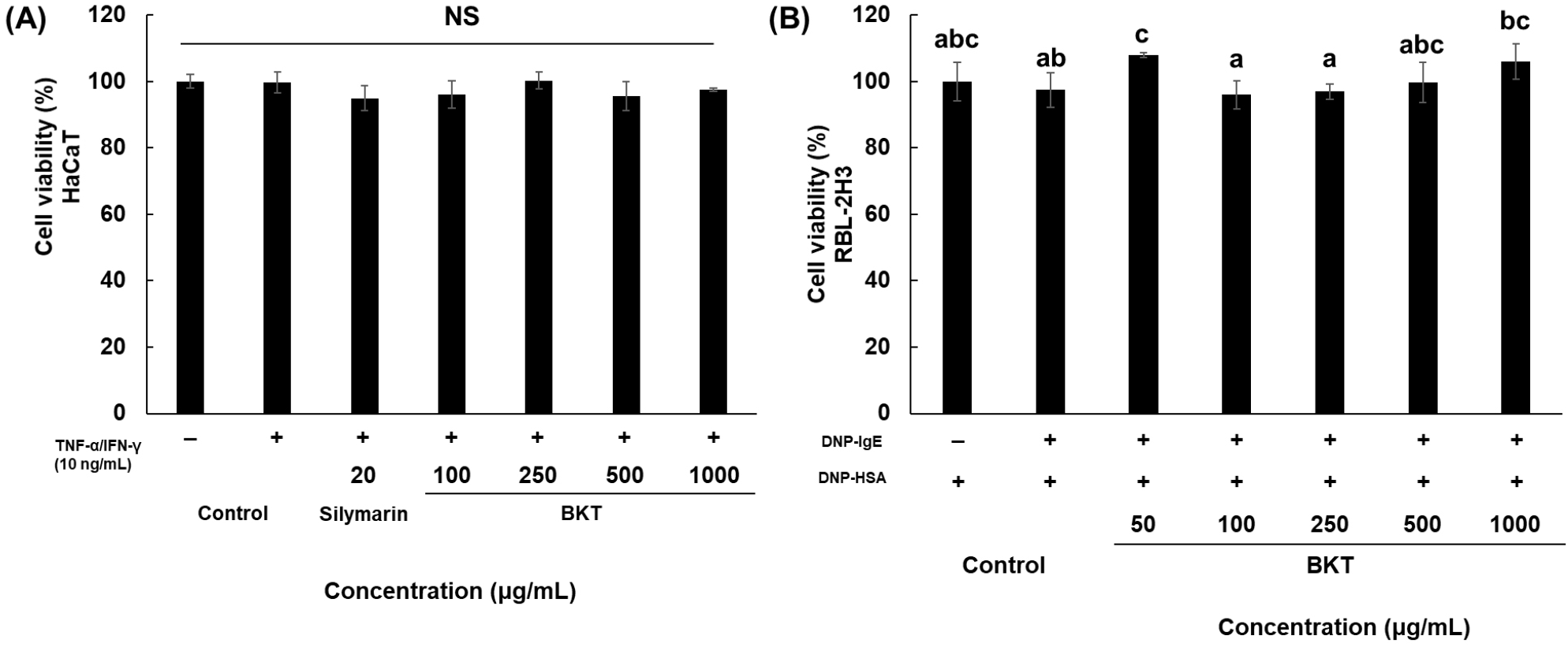
HaCaT 피부각질세포에 독성을 나타내지 않는 닥나무 가지 추출물의 농도를 결정하기 위하여 MTT assay를 이용한 세포생존율을 확인하였다. HaCaT 피부각질세포에 10 ng/mL T+I와 시료를 100-1,000 μg/mL 농도로 처리한 결과(Fig. 1A), 닥나무 가지 추출물은 1,000 μg/mL 농도까지 대조군 대비 세포독성이 나타나지 않았다.

kjfp-31-2-307-g1
Fig. 1. Cytotoxicity of Broussonetia kazinoki Twig extract (BKT) on (A) HaCaT and (B) RBL-2H3 cells. Cell viability was measured by MTT assay. Values are mean±SD (n=3). Different letters (a-c) on the bar indicate significant differences (p<0.05). NS, not significant.
Download Original Figure
3.2. 염증성 케모카인 생성 억제 효과

닥나무 가지 추출물의 HaCaT 세포에 대한 TARC, MDC, RANTES 생성에 미치는 영향을 평가하였다. 10 ng/mL T+I를 처리하여 염증을 유도한 HaCaT 피부각질세포에 대한 염증성 케모카인 생성 억제 효과를 측정한 결과는 Fig. 2와 같다. 본 실험에서 10 ng/mL T+I 처리를 통해 HaCaT 피부각질세포 내의 TARC, MDC, RANTES 생성이 증가되었으며(Fig. 2A-2C), 100, 250, 500, 1,000 μg/mL의 닥나무 가지 추출물을 처리하였을 때 TARC, MDC, RANTES는 대조군 대비 염증성 케모카인 생성 억제율이 각각 최대 60.45%, 50.94%, 69.65%로, positive control인 silymarin보다 더 높은 억제활성을 나타내었으며, 농도의존적인 염증성 케모카인 생성 억제를 나타내었다.

kjfp-31-2-307-g2
Fig. 2. Effect of Broussonetia kazinoki Twig extract (BKT) on (A) TARC, (B) MDC and (C) RANTES production in TNF-α/IFN-γ stimulated HaCaT cells. HaCaT cells were co-treated with BKT and TNF-α + IFN-γ for 24 h. After treatment, the production levels of TARC, MDC and RANTES were determined in cell cultured media and using a commercial ELISA kit. Values are mean±SD (n=3). Different letters (a-f) on the bar indicate significant differences (p<0.05).
Download Original Figure

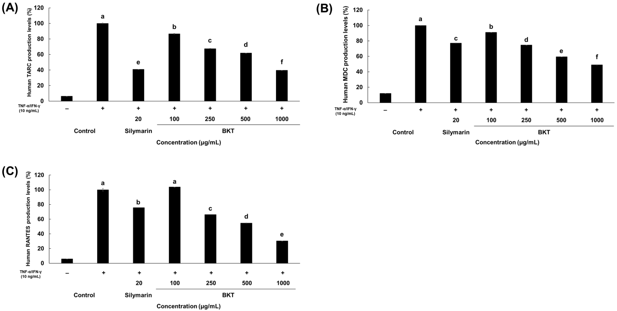
인간유래 각질형성 세포주인 HaCaT 세포는 염증성 사이토카인(TNF-α 및 IFN-γ, T+I) 처리 시 IL-6와 같은 사이토카인, IL-8, TARC, MDC 및 RANTES와 같은 케모카인 분비를 촉진하여 AD를 유발하는 것으로 알려져 있다(Eom 등, 2022; Ha 등, 2020). 또한, 신체에서 피부 장벽이 손상되어 항원이 체내에 침투하면 각질형성세포는 TSLP를 분비하여 수지상 세포를 자극해 TARC와 같은 케모카인의 생성을 증가시킨다(Lee, 2010). 이러한 케모카인은 알러지 염증, AD, 알러지성 비염 등에서 유의하게 증가하는 지표로서 호산구, 비만세포, Th2 세포들을 염증 부위로 이동시키는 역할을 한다(Sandoval‐Lopez과 Teran, 2001). 염증성 케모카인은 염증반응을 매개하는 인자로 작용하여 피부염증 등의 유발인자가 될 수 있으므로 피부염증 관련 질병의 억제와 치료를 위해서는 이러한 염증성 케모카인의 생성이 억제되어야 한다.

닥나무 가지 추출물이 3가지 케모카인 TARC, MDC, RANTES 생성에 미치는 영향을 확인한 결과, 10 ng/mL의 T+I에 의해 증가된 TARC, MDC, RANTES 생성을 유의하게 억제하였다. 본 실험결과는 닥나무 가지가 피부각질세포에서 케모카인 분비를 억제하여 알러지성 피부 염증 반응을 효과적으로 개선할 수 있는 소재로 사용될 수 있음을 시사한다.

3.3. 닥나무 가지 추출물의 RBL-2H3 세포에 대한 세포 독성

RBL-2H3 세포에 독성을 나타내지 않는 닥나무 가지 추출물의 농도를 결정하기 위하여 MTT assay를 이용한 세포생존율을 확인하였다. RBL-2H3 세포를 DNP-IgE로 감작한 후 시료를 100-1,000 μg/mL 농도로 처리한 결과(Fig. 1B), 닥나무 가지 추출물은 1,000 μg/mL 농도까지 대조군 대비 세포독성이 나타나지 않았다.

3.4. 닥나무 가지 추출물이 β-hexosaminidase 방출에 미치는 영향

DNP-IgE에 의해 감작된 RBL-2H3 세포에서 닥나무 가지 추출물 100, 250, 500, 1,000 μg/mL 농도 처리가 β-hexosaminidase 방출에 미치는 영향은 Fig. 3과 같다. RBL-2H3 cell에 DNP-IgE 감작 후 DNP-HSA 처리는 β-hexosaminidase 방출을 증가시켰고, 증가된 β-hexosaminidase는 닥나무 가지 추출물에 의해 농도의존적으로 감소하였으며, 닥나무 가지 추출물 100, 250, 500, 1,000 μg/mL 농도에서 각각 2.70%, 6.50%, 54.25%, 98.35% 감소하였다.

kjfp-31-2-307-g3
Fig. 3. The inhibitory effect of Broussonetia kazinoki Twig extract (BKT) on β-hexosaminidase release in IgE-sensitized RBL-2H3 cells. IgE-sensitized RBL-2H3 cells were pretreated with BKT for 1 h prior to exposure to DNP-HSA for 4 h. Values are mean±SD (n=3). Different letters (a-c) on the bar indicate significant differences (p<0.05).
Download Original Figure

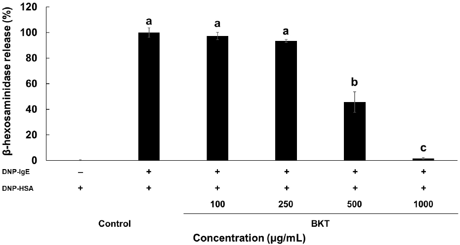
비만세포는 알러지의 주요인이 되는 면역세포로서, 비만세포라고 불리는 이유는 핵에 비해 세포질이 많고 세포질 내에 과립을 많이 갖고 있기 때문이다. 따라서 알러지 비만세포 모델로 호염기 세포(basophils) RBL-2H3 세포가 많이 사용되고 있다(Chen 등, 2012; Passante와 Frankish, 2009). 또한, 이들 비만세포가 활성화되었을 때 분비되는 히스타민 농도가 낮아 히스타민과 함께 분비되는 β-hexosaminidase 활성을 측정함으로써 탈과립 정도를 알 수 있다(Chung 등, 2011; Chung 등, 2013; Hwang 등, 2018). 세포의 탈과립 정도를 알 수 있는 β-hexosaminidase 방출 억제 효과를 가진 천연식품소재 발굴은 알러지 반응을 유발할 수 있는 식품으로 개발할 때 알러지를 저감화할 수 있는 제품에 활용할 수 있을 것이며, 건강기능식품, 건강보조식품 개발 등 광범위한 분야에 활용할 수 있을 것이다. 본 연구에서 사용한 닥나무 가지 추출물은 β-hexosaminidase 방출을 억제함으로써 항알러지 효과가 있음을 알 수 있다.

3.5. 닥나무 가지 추출물이 TNF-α, IL-4 생성에 미치는 영향

닥나무 가지 추출물을 DNP-IgE에 의해 감작된 RBL-2H3 세포에 처리한 결과, TNF-α, IL-4 발현에 미치는 영향은 Fig. 4와 같다. RBL-2H3 cell에 DNP-IgE 감작 후 DNP-HSA 처리는 TNF-α, IL-4의 방출을 증가시켰고, 증가된 TNF-α, IL-4는 닥나무 가지 추출물 250, 500 그리고 1,000 μg/mL 농도 처리에 의해 감소하였으며, 닥나무 가지 추출물 100, 250, 500, 1,000 μg/mL 농도에서 TNF-α가 각각 22.52, 49.23, 89.10, 98.13% 감소하였고, IL-4는 6.03, 63.35, 89.18, 89.22% 감소하였다.

kjfp-31-2-307-g4
Fig. 4. Effect of Broussonetia kazinoki Twig extract (BKT) on (A) TNF-α and (B) IL-4 production levels in IgE-sensitized RBL-2H3 cells. IgE-sensitized RBL-2H3 cells were pretreated with BKT for 1 h prior to exposure to DNP-HSA for 4 h. Values are mean±SD (n=3). Different letters (a-d) on the bar indicate significant differences (p<0.05). TNF-α and IL-4 production levels were determined in cell cultured media and using a commercial ELISA kit.
Download Original Figure

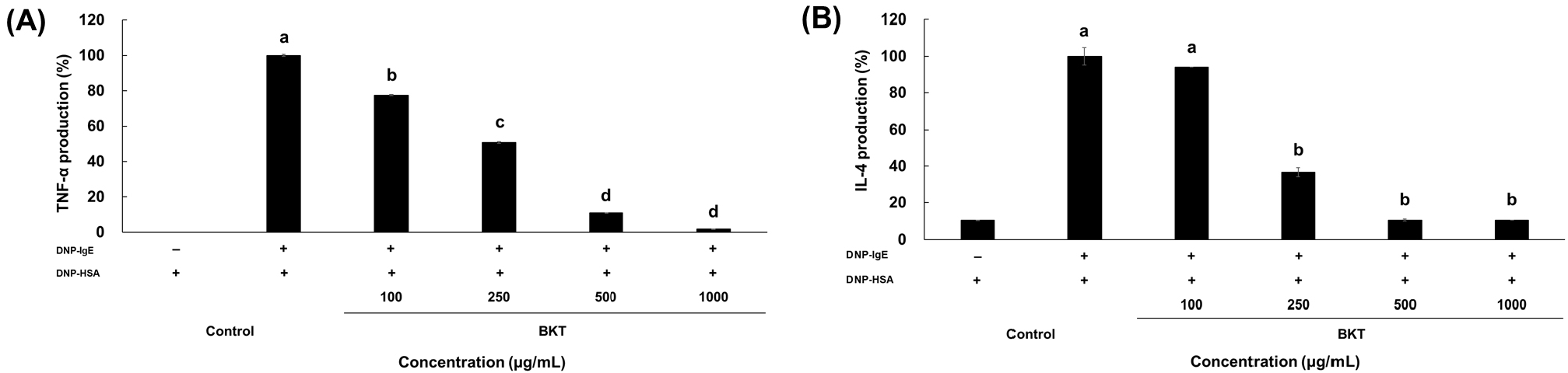
비만세포가 활성화되면 TNF-α와 IL-4의 발현과 분비가 증가되는 것으로 알려져 있다(Lee 등, 2023). 안토시아닌을 포함한 플라보노이드 등 다양한 소재들이 알러지 세포모델 또는 동물모델에서 사이토카인 생성을 억제함으로써 알러지 관련 질환을 완화시킬 수 있을 것이라는 보고들이 있고(Chung 등, 2011), 석곡 열수 추출물 처리가 탈과립 및 알러지 관련 사이토카인(TNF-α 그리고 IL-4)의 생성을 감소함으로써 항알러지 효과가 있다고 보고하였다(Lee 등, 2023).

닥나무는 전통적으로 종이의 재료, 염증성 질환 및 전염병 등을 치료하는 약으로 쓰여왔다. 또한, 닥나무는 이소프레닐화(isoprenylated) 및 프레닐화(prenylated) 플라반(flavan), Broussoneinine M, O, P와 같은 생리활성 화합물을 포함하는 것으로 알려져 있다(Cho 등, 2014; Shibano 등, 1997). 닥나무를 포함한 많은 약용 식물에는 항염증 및 항산화 효능이 있는 탄닌과 플라보노이드를 포함한 페놀성 화합물이 포함되어 있으며(Cho 등, 2014; Diaz 등, 2012), 닥나무는 항당뇨, 항고혈당, 티로시나제 및 멜라닌 합성 억제 등(Cha 등, 2008; Hwang과 Lee, 2007; Ryu 등, 2003)의 여러 효능이 보고되었다. 이에 본 연구에서는 닥나무 가지가 피부질환 관련 각질형성세포인 HaCaT 세포와 호염기성 백혈병 비만세포인 RBL-2H3 세포에서 케모카인, β-hexosaminidase 방출, 염증성 사이토카인의 분비에 대한 영향을 분석하였다.

본 연구 결과, 닥나무 가지 추출물이 비만세포가 활성화될 때 분비되는 β-hexosaminidase 및 사이토카인(TNF-α 그리고 IL-4)의 발현을 현저하게 억제하였으므로 비만세포로 인한 알러지 반응 또는 염증반응을 완화하여 알러지 저감화 효과를 유도할 수 있을 것으로 사료된다.

4. 요약

닥나무는 항산화, 항암 효과뿐 아니라 천연 미백기능성분으로 인정받아 화장품 원료로 사용되며 닥나무의 잎과 가지는 식약처에 등재된 식용이 가능한 원료이다. 닥나무는 아시아에서 종이 제조 및 한의학적 용도로 사용되었고 항당뇨 등의 효능이 있으며, 다양한 플라보노이드와 알칼로이드를 포함하는 것으로 알려져 있다. 본 연구에서는 닥나무 추출물의 피부 염증성 알러지 반응에 대한 효능을 평가하기 위하여, HaCaT 각질형성세포의 피부염증 억제와 RBL-2H3 세포의 알러지 반응 억제와 관련된 인자들에 미치는 영향에 관하여 연구하였다. HaCaT 및 RBL-2H3 세포에 대한 70% 에탄올 추출물의 세포독성은 나타나지 않았다. HaCaT 세포에서 TNF-α와 IFN-γ의 자극으로 케모카인(TARC, MDC, RANTES) 생성이 증가하였으며, 시료 처리에 따라 농도의존적으로 유의성 있게 감소하였다. 한편, IgE 처리로 활성화된 RBL-2H3 세포에서 증가하는 β-hexosaminidase 방출과 염증성 사이토카인 TNF-α, IL-4 생성이 시료 처리로 유의적으로 감소하는 것을 확인하였다. 따라서 닥나무 가지 추출물은 알러지 염증반응 완화 효과를 갖는 천연물 화장품 및 식품 원료로 활용이 가능할 것으로 예상된다.

Acknowledgements

None.

Conflict of interests

The authors declare no potential conflicts of interest.

Author contributions

Conceptualization: Yang SA. Methodology: Bae WB, Yang SA. Formal analysis: Bae WB, Kim EH. Validation: Bae WB, Kim MJ. Writing - original draft: Bae WB, Kim EH. Writing - review & editing: Bae WB, Yang SA.

Ethics approval

This article does not require IRB/IACUC approval because there are no human and animal participants.

Funding

This research was supported by Basic Science Research Program through the National Research Foundation of Korea (NRF) funded by the Ministry of Education (NRF-2021R1I1A3055952).

ORCID

Won-Bin Bae (First author) https:orcid.org/0009-0009-5733-5972

Eun-Hye Kim https:orcid.org/0009-0005-0793-1259

Min-Ju Kim https:orcid.org/0009-0005-7918-9428

Seun-Ah Yang (First & Corresponding author) https:orcid.org/0000-0002-6692-1844

References

1.

Bergmann RL, Edenharter G, Bergmann KE, Forster J, Bauer CP, Wahn V, Zepp F, Wahn U. Atopic dermatitis in early infancy predicts allergic airway disease at 5 years. Clin Exp Allergy. 28:965-970 1998;

2.

Cha JY, Kim YT, Kim HS, Cho YS. Antihyperglycemic effect of stem bark powder from paper mulberry (Broussonetia kazinoki Sieb.) in type 2 diabetic otsuka long-evans tokushima fatty rats. J Med Food. 11:499-505 2008;

3.

Chen BH, Hung MH, Chen JYF, Chang HW, Yu ML, Wan L, Tsai FJ, Wang TP, Fu TF, Chiu CC. Anti-allergic activity of grapeseed extract (GSE) on RBL-2H3 mast cells. Food Chem. 132:968-974 2012;

4.

Cho YR, Kim JH, Kim JK, Ahn EK, Ko HJ, In JK, Lee SJ, Bae GU, Kim YK, Oh JS. Broussonetia kazinoki modulates the expression of VEGFR-2 and MMP-2 through the inhibition of ERK, AKT and p70s6k-dependent signaling pathways: Its implication in endothelial cell proliferation, migration and tubular formation. Oncol Rep. 32:1531-1536 2014;

5.

Chung MJ, Ha TJ, Choi HN, Lee JS, Par YI. Inhibitory effects of anthocyanins isolated from black soybean (Glycine max L.) seed coat on degranulation and cytokine generation in RBL-2H3 cells. J Korean Soc Food Sci. 40:1662-1667 2011;

6.

Chung MJ, Sohng JK, Choi DJ, Park YI. Inhibitory effect of phloretin and biochanin a on IgE-mediated allergic responses in rat basophilic leukemia RBL-2H3 cells. Life Sci. 93:401-408 2013;

7.

Diaz P, Jeong SC, Lee S, Khoo C, Koyyalamudi SR. Antioxidant and anti-inflammatory activities of selected medicinal plants and fungi containing phenolic and flavonoid compounds. Chin Med. 7:1-9 2012;

8.

Eom HJ, Shin HY, Park HJ, Kim KH, Kim JH, Yu KW. Functional components and physiological activity in different parts of Centella asiatica. Korean J Food Preserv. 29:749-761 2022;

9.

Furukawa H, Nakamura K, Zheng X, Tojo M, Oyama N, Akiba H, Nishibu A, Kaneko F, Tsunemi Y, Saeki H, Tamaki K. Enhanced TARC production by dust-mite allergens and its modulation by immunosuppressive drugs in PBMCs from patients with atopic dermatitis. J Dermatol Sci. 35:35-42 2004;

10.

Ha Y, Lee WH, Jeong J, Park M, Ko JY, Kwon OW, Lee J, Kim YJ. Pyropia yezoensis extract suppresses IFN-gamma- and TNF-alpha-induced proinflammatory chemokine production in HaCaT cells via the down-regulation of NF-κb. Nutrients. 12:1238 2020;

11.

Hashimoto S, Nakamura K, Oyama N, Kaneko F, Tsunemi Y, Saeki H, Tamaki K. Macrophage-derived chemokine (MDC)/CCL22 produced by monocyte derived dendritic cells reflects the disease activity in patients with atopic dermatitis. J Dermatol Sci. 44:93-99 2006;

12.

Homey B, Zlotnik A. Chemokines in allergy. Curr Opin Immunol. 11:626-634 1999;

13.

Hudson TJ. Skin barrier function and allergic risk. Nat Genet. 38:399-400 2006;

14.

Hwang JH, Lee BM. Inhibitory effects of plant extracts on tyrosinase, L-dopa oxidation, and melanin synthesis. J Toxicol Environ Health Part A. 70:393-407 2007;

15.

Hwang KA, Hwang YJ, Song J. Anti-allergic effect of aster yomena on ovalbumin-sensitized mouse and RBL-2H3 cells via Th1/Th2 cytokine balance. J Funct Foods. 44:1-8 2018;

16.

Itoh T, Ohguchi K, Nakajima C, Oyama M, Iinuma M, Nozawa Y, Akao Y, Ito M. Inhibitory effects of flavonoid glycosides isolated from the peel of Japanese persimmon (Diospyros kaki Fuyu) on antigen-stimulated degranulation in rat basophilic leukaemia RBL-2H3 cells. Food Chem. 126:289-294 2011;

17.

Jo IJ. Inhibitory activity of terminalia chebula extract against TNF-α/IFN-γ-induced chemokine increase on human keratinocyte, HaCaT cells. Kor J Herbology. 37:41-47 2022;

18.

Kim MJ, Hwang BS, Hwang Y, Jeong YT, Jeong DW, Oh YT. Anti-Inflammatory and antiatopic effects of Rorippa cantoniensis (Lour.) Ohwi in RAW 264.7 and HaCaT cells. Molecules. 28:5463 2023;

19.

Lee A. Immunologic barrier in skin of atopic dermatitis. Atopic Dermat Symp. p. S143-S148 2010;

20.

Lee JK, Ha H, Lee HY, Park SJ, Jeong SL, Choi YJ, Shin HK. Inhibitory effects of heartwood extracts of Broussonetia kazinoki Sieb on the development of atopic dermatitis in Nc/Nga mice. Biosci Biotechnol Biochem. 74:1802-1806 2010;

21.

Lee JY, Park SH, Jhee KH, Yang SA. Tricin isolated from enzyme-treated Zizania latifolia extract inhibits IgE-mediated allergic reactions in RBL-2H3 cells by targeting the Lyn/Syk pathway. Molecules. 25:2084 2020;

22.

Lee N, Ha H, Lee H, Jung D, Choi Y, Jeong S, Shin H. Analysis of studies on the by-products of the Broussonetia kazinoki Siebold or Broussonetia papyrifera (L.) Vent. Korean J Ori Med. 14:49-58 2008;

23.

Lee YJ, Madhi I, Kim Y. Inhibitory effect of Dendrobium moniliforme on degranulation and histidine decarboxylase expression in RBL-2H3 cells. J Life Sci. 33:176-182 2023;

24.

Oh JS, Jang HH. Epidermal differentiation and skin barrier. Kor J Aesthet Cosmetol. 13:713-720 2015;

25.

Passante E, Frankish N. The RBL-2H3 cell line: Its provenance and suitability as a model for the mast cell. Inflamm Res. 58:737-745 2009;

26.

Ryu JH, Ahn H, Jin Lee H. Inhibition of nitric oxide production on LPS-activated macrophages by kazinol B from Broussonetia kazinoki. Fitoterapia. 74:350-354 2003;

27.

Sandoval-Lopez G, Teran L. TARC: Novel mediator of allergic inflammation. Wiley Online Library, Clin Exp Allergy. 31:1809-1812 2001;

28.

Schroder JM, Noso N, Sticherling M, Christophers E. Role of eosinophil-chemotactic CC chemokines in cutaneous inflammation. J Leucoc Biol. 59:51-55 1996;

29.

Shibano M, Kitagawa S, Nakamura S, Akazawa N, Kusano G. Studies on the constituents of Broussonetia species. Ii. Six new pyrrolidine alkaloids, broussonetine a, b, e, f and broussonetinine a and b, as inhibitors of glycosidases from Broussonetia kazinoki Sieb. Chem Pharm Bull. 45:700-705 1997;

30.

Sicherer SH, Sampson HA. Food allergy: A review and update on epidemiology, pathogenesis, diagnosis, prevention, and management. J Allergy Clin Immunol. 141:41-58 2018;

31.

Tsukamoto D, Shibano M, Kusano G. Studies on the constituents of Broussonetia species x. Six new alkaloids from Broussonetia kazinoki sieb. Chem Pharm Bull. 49:1487-1491 2001;

32.

Zhang PC, Wang S, Wu Y, Chen RY, Yu DQ. Five new diprenylated flavonols from the leaves of Broussonetia kazinoki. J Nat Prod. 64:1206-1209 2001;